Reduction of a large thoracic scoliosis using a ScoliBrace in a young ballerina
Summary:
This case details the reduction of a moderate-severe thoracic scoliosis in a 17-year-old female ballet dancer using a ScoliBrace orthosis combined with scoliosis specific rehabilitation. The patient presented at the end of her adolescent growth phase with a 44° (Cobb) thoracic scoliosis with associated coronal imbalance. The patient was also experiencing frequent bouts of lower back pain.
The patient was prescribed a rigid ScoliBrace to be worn on a part-time basis and advised to perform daily rehabilitation exercises. The patient wore the brace as prescribed for 12 months. At the end of treatment, the patient’s curve had been reduced from 44° down to 29° (Cobb) and the patient reported that her pain levels had reduced dramatically.
See More: ScoliBrace Reviews: Reduction of scoliosis 14 old patient
Case Background
The 17-year-old female patient presented to the ScoliCare clinic with scoliosis and lower back pain. The patient had been diagnosed with scoliosis 18 months prior by a general practitioner.
The patient was a ballet dancer, attending classes full-time, with aspirations to pursue dancing as a career. The patient’s scoliosis was causing significant postural asymmetries and obvious rib humping. The patient and the parents were concerned that the deformity and pain caused by the scoliosis may interfere with the patient’s ballet career.
Furthermore, the patient did not want to undergo surgery as she felt that this would impact negatively on her flexibility and movement. Instead, the patient was looking for a more conservative type of treatment to help reduce her scoliosis and to alleviate her pain.
Examination Findings
The patient reported that she was experiencing lower back pain most days measuring 7-8/10 on a visual analog scale (VAS). The patient’s medical history, family history, and systems review were unremarkable.
The physical examination revealed a prominent curvature in the spine, waist curve asymmetry, scapula malpositioning and significant right coronal imbalance (Figure 1 [Left]). Rib humping was apparent during the Adams forward bend test. The patient’s lower back pain was reproduced when the Kemps test was performed in the lumbar spine on the right.
No abnormalities were detected during the neurological examination.
X-rays taken at the time of the initial consultation demonstrated that the patient had a 44° (Cobb) left thoracic scoliosis with an apex at T10-11 intervertebral disc (Figure 1 [Right]). The patient was Risser 5 at the time of the initial consultation.
A modified type of bending x-ray was used to assess the patients curve flexibility. To perform this assessment the patient is taught how to perform an active self-correction. In this position, the patient is then x-rayed to determine the potential for a correction. As can be seen in (Figure 2) the patient’s curve shows good flexibility reducing to 23° in the active self-corrected position.
The patient reported that she had also consulted with an orthopedic surgeon. The surgeon had performed a physical examination and ordered an MRI. Aside from thoracic scoliosis, there were no other abnormalities detected on the MRI. The surgeon had advised the patient that, given her age and the magnitude of the curve, surgery was the most suitable treatment option for her scoliosis.
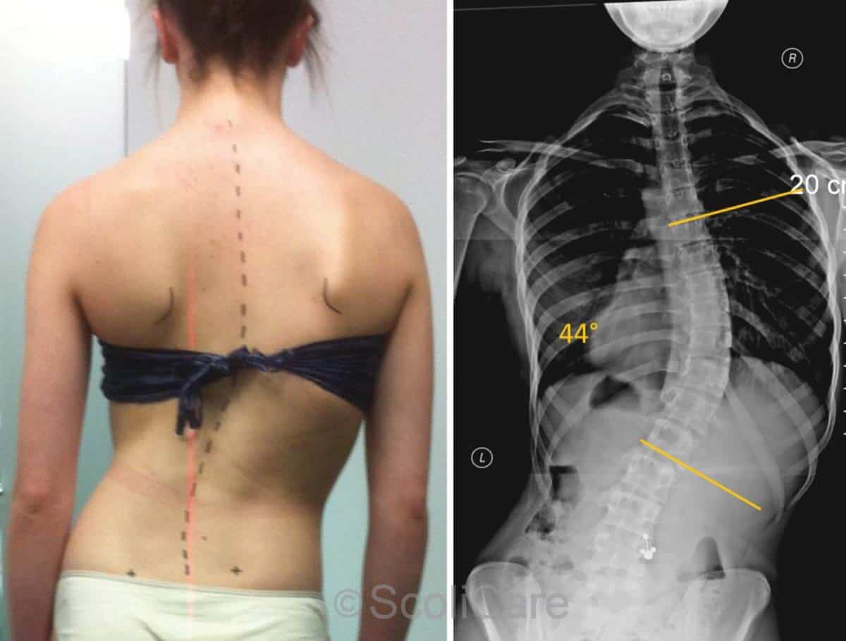
Figure 1: Posteroanterior postural photograph (left), posteroanterior full-spine radiograph (right)
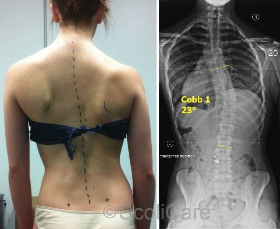
Figure 2: Posteroanterior photograph with the patient
performing an active self-correction (Left)
Intervention
The patient and her parents had refused surgery and had made the decision to trial conservative management.
The patient was subsequently prescribed a ScoliBrace, rigid thoracolumbosacral orthosis [TLSO] (Figure 3). As the patient was attending daily ballet classes, the patient was advised to wear the brace on a part-time basis (6-8 hours per day) for 6-12 months.
The patient was also prescribed daily scoliosis specific exercise to complement the action of the TLSO. The patient was advised to perform the rehabilitation exercises twice daily for approximately 20 minutes.
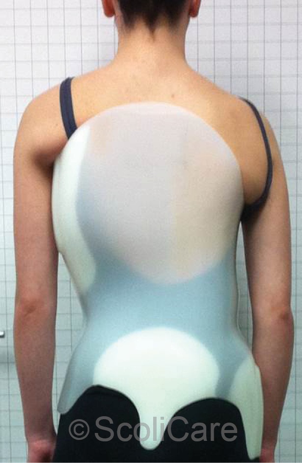
Figure 3: Posteroanterior photograph of the patient wearing the custom made TLSO
Outcomes
The patient was compliant with brace wear and the rehabilitation program. After six months of treatment there were noticeable changes in the patient’s posture. Specifically, a reduction in the cosmetic deformity and coronal balance was observed (Figure 4).
The patient continued with the bracing and rehabilitation exercises for a further six months. By the end of the 12-month treatment period the patient reported that there had been a significant reduction in the pain that she had been experiencing.
The patient stated that she only experienced minor pain (2-3/10 [VAS]) on occasion, compared with the strong pain that she was experiencing on a daily basis prior to commencing the treatment. Postural photographs taken at the end of the treatment period highlighted that the patient had remained balanced in the coronal plane, there was a reduction in the asymmetry of the waist curves, decreased rib humping and an associated improvement in the scapula positioning (Figure 4 [Left]).
X-rays taken at an independent radiology practice revealed that the patient’s scoliosis had reduced by 15° (Cobb) down to 29° in response to the bracing and exercise regimens (Figure 5 [Right]).
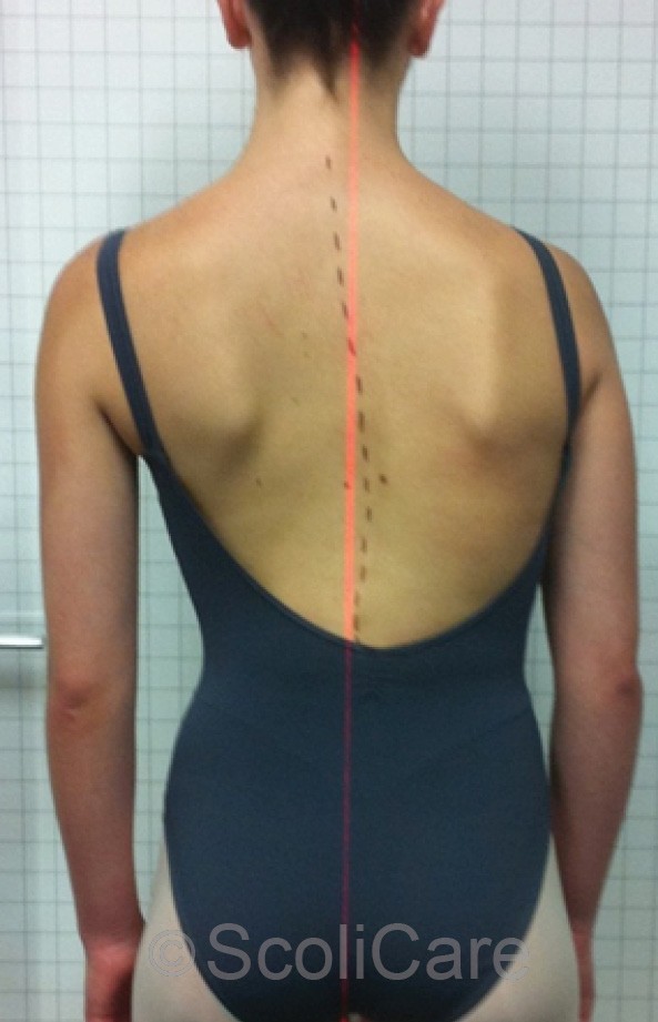
Figure 4: Posteroanterior postural photograph taken after six months of treatment.
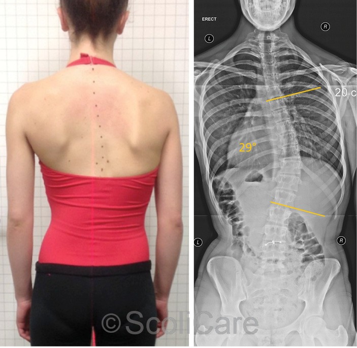
Figure 5: Post-treatment posteroanterior postural photograph (Left) Post-treatment posteroanterior x-rays demonstrating a left thoracic curve measuring 29° (Cobb).
Discussion
This case demonstrates the successful management of a skeletally mature patient with a moderate-severe thoracic scoliosis.
There is mounting evidence that children and adolescents experience acute and chronic back pain, which often continues into adulthood. Pain is amplified in patients who are involved with demanding physical activities such as elite level sport.
The presence of scoliosis further complicates the clinical scenario for these individuals creating abnormal loading and biomechanical stresses on the spinal column. In this case, the patient was involved in intensive ballet training for prolonged periods each day.
This combined with a large magnitude curve with an associated coronal imbalance was likely contributing to the patient’s pain. The chances of future curve progression were also quite high in this case given the initial magnitude of the patient’s scoliosis and her high levels of physical activity, and the fact that the patient was coronally imbalanced.
Despite being skeletally mature the good flexibility observed in the active self-correction x-ray suggested that there was potential for the curve to be improved.
Using bracing and scoliosis specific exercises this patient was able to avoid surgery, dramatically improve her quality of life and reduce the cosmesis associated with her scoliosis.
The patient was selected to join a top ballet institution overseas and was advised to continue with the scoliosis specific exercises to stabilise the correction that had been achieved and counteract the high physical demands associated with the ballet training.
Conclusion
This case demonstrates the ScoliBrace orthosis combined with scoliosis specific exercises can be used to manage some skeletally mature patients with thoracic curves that have reached the surgical threshold where that curve is unbalanced but remains relatively flexible.
In this case, a significant improvement in aesthetics, curve magnitude, coronal balance and pain were achieved using this conservative approach.
NB: Results vary from case to case. Our commitment is to recommend the most appropriate treatment based on the patients type and severity of scoliosis.
ScoliCare, Australia
